Technology hero banner

Technology

Technology hero banner

Technology

Technology hero banner

Technology

Technology hero banner

Technology

Circulating Tumor Cells (CTCs)?

The Invisible Threat

The Invisible Threat

What Are Circulating Tumor Cells (CTCs)?

What Are Circulating Tumor Cells (CTCs)?

CTCs are cancer cells that detach from primary or metastatic tumors and enter the bloodstream. They carry the same genetic and molecular information as the tumor itself — a “liquid window” into cancer’s evolution. By analyzing CTCs, clinicians can monitor disease progression, treatment response, and emerging resistance, all from a simple blood draw.

CTCs are cancer cells that detach from primary or metastatic tumors and enter the bloodstream. They carry the same genetic and molecular information as the tumor itself — a “liquid window” into cancer’s evolution. By analyzing CTCs, clinicians can monitor disease progression, treatment response, and emerging resistance, all from a simple blood draw.

The Challenge of

The Challenge of

Detecting Circulating Tumor Cells

Detecting Circulating Tumor Cells

Circulating tumor cells (CTCs) are extraordinarily rare and heterogeneous, making their detection one of the most demanding tasks in cancer diagnostics. They can differ in size, shape, and molecular profile, and often exist as single cells or small clusters hidden among billions of normal blood cells. Traditional detection methods struggle to capture this diversity, leading to inconsistent results and missed opportunities for early intervention. Understanding and overcoming these challenges is essential to unlocking the full potential of liquid biopsy for cancer management.

Circulating tumor cells (CTCs) are extraordinarily rare and heterogeneous, making their detection one of the most demanding tasks in cancer diagnostics. They can differ in size, shape, and molecular profile, and often exist as single cells or small clusters hidden among billions of normal blood cells. Traditional detection methods struggle to capture this diversity, leading to inconsistent results and missed opportunities for early intervention. Understanding and overcoming these challenges is essential to unlocking the full potential of liquid biopsy for cancer management.

Detecting Circulating Tumor Cells

How We Overcame the Challenge

How We Overcame the Challenge

The Labyrinth One Microfluidic System

The Labyrinth One Microfluidic System

At BloodScan, we turned to fluid dynamics to solve a biological problem. Our patented Labyrinth microfluidic chip uses precise channel geometry to control inertial flow and Dean vortices — hydrodynamic effects that naturally separate cells by size, shape, and deformability. Without labels or antibodies, we isolate intact and viable CTCs at unmatched purity and yield.

At BloodScan, we turned to fluid dynamics to solve a biological problem. Our patented Labyrinth microfluidic chip uses precise channel geometry to control inertial flow and Dean vortices — hydrodynamic effects that naturally separate cells by size, shape, and deformability. Without labels or antibodies, we isolate intact and viable CTCs at unmatched purity and yield.

The Labyrinth One Microfluidic System

From blood draw to purified CTCs in minutes

From blood draw to purified CTCs in minutes

Watch how Labyrinth One transforms routine blood samples into high-quality circulating tumor cells through a fully automated, plug-and-play workflow.

Automated, label-free microfluidic separation

High throughput and clinical scalability

Preserves live, intact cells for downstream assays

How It Works

How It Works

STEP 1
Blood In, Biology Out

A standard blood draw — just 7–10 mL — is all it takes. Within minutes, the sample is processed through the Labyrinth One microfluidic cartridge, no labels or reagents required.
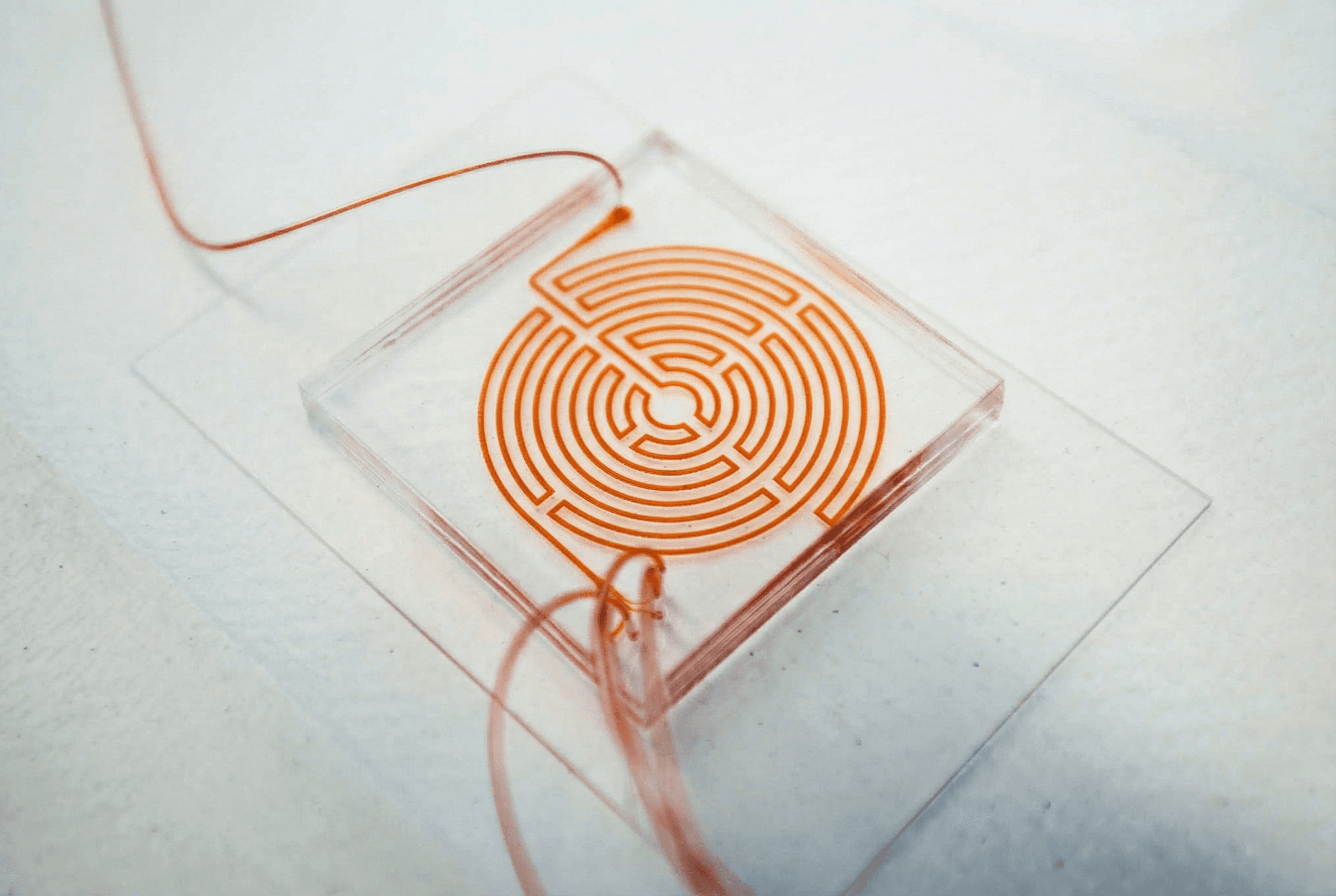
STEP 2
Inside the Labyrinth

Our patented Labyrinth microfluidic chip uses intricate channel geometry to create precise fluidic dynamics. These natural flow patterns separate circulating tumor cells (CTCs) from billions of normal blood cells — purely based on size, shape, and deformability.

STEP 3
Isolate, Identify, and Analyze

The isolated CTCs remain intact and viable, ready for a wide range of analyses —
from immunostaining and FISH to single-cell sequencing, organoid culture, and drug testing.

Proven Performance

Proven Performance

BloodScan’s technology builds on over a decade of research at the University of Michigan, validated across leading cancer centers worldwide. Our results demonstrate that liquid biopsy can now achieve tissue-level insight — non-invasively, repeatedly, and in real time.

BloodScan’s technology builds on over a decade of research at the University of Michigan, validated across leading cancer centers worldwide. Our results demonstrate that liquid biopsy can now achieve tissue-level insight — non-invasively, repeatedly, and in real time.

Early Detection

CTCs detected as early as stage 0 (DCIS, early HCC)

Sensitivity

100 % capture in late-stage cancers (breast, NSCLC, pancreatic, and more)

Genomic Concordance

> 97 % similarity between CTCs and matched tissue biopsies

Cell Viability

Intact, ready for histology, genomics, or advanced assays

Publications

Supported by > 15 peer-reviewed papers worldwide

Early Detection

CTCs detected as early as stage 0 (DCIS, early HCC)

Cell Viability

Intact, ready for histology, genomics, or advanced assays

Sensitivity

100 % capture in late-stage cancers (breast, NSCLC, pancreatic, and more)

Publications

Supported by > 15 peer-reviewed papers worldwide

Genomic Concordance

> 97 % similarity between CTCs and matched tissue biopsies